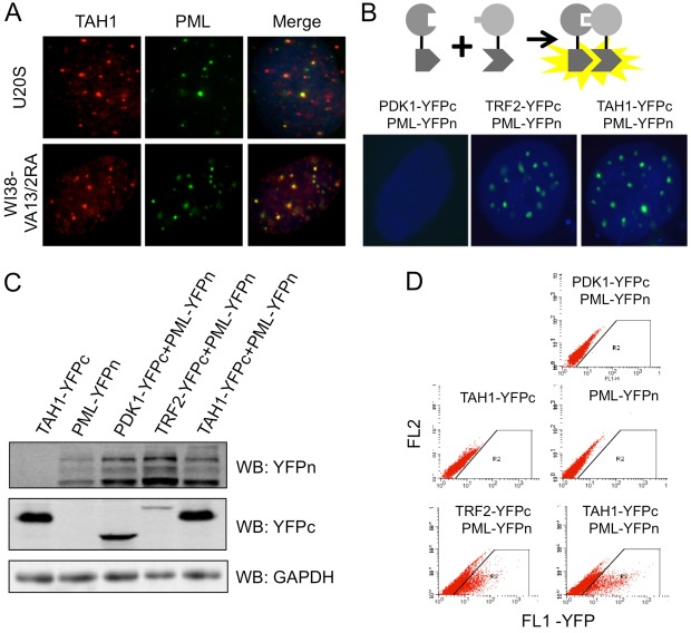
Fig. 3.
TAH1 localizes to PML bodies in ALT cells. (A) U2OS cells and WI38-VA13/2RA cells were co-immunostained with antibodies against endogenous TAH1 (red) and PML (green). (B) BiFC assays were carried out in U2OS cells stably expressing PML-YFPn plus TAH1-YFPc. Cells expressing PML-YFPn plus PDK1-YFPc or PML-YFPn plus TFR2-YFPc were used as negative or positive controls, respectively. (C) Western blotting for protein expression in the indicated cell lines. (D) FACS analysis of fluorescence complementation in cells from C. U2OS cells expressing TAH1-YFPc only, PML-YFPn only, and PML-YFPn plus PDK1-YFPc were used as negative controls.

