
Fig. 6.
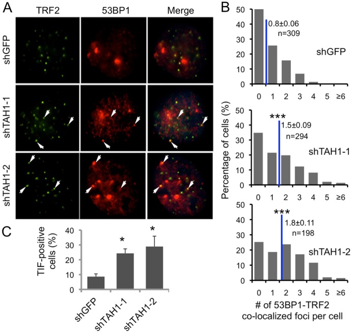
TAH1 regulates telomere DNA damage responses in ALT cells. (A) Representative images of the telomere-dysfunction-induced foci (TIF) analysis in control and TAH1 knockdown U2OS cells using anti-53BP1 (red) and TRF2 (green) antibodies. Arrows indicate superimposable foci. (B) 53BP1 and TRF2 co-staining foci (TIFs) were scored for individual cells from A. Blue lines mark the average number of TIFs per cell. Error bars indicate standard errors (n = 3). ***P<0.001 compared with control. (C) Percentages of TIF-positive cells were calculated based on data from A and B. Cells with ≧3 co-localization foci were scored as TIF positive. Error bars indicate standard errors (n = 3). *P<0.05 compared with control.