
Fig. 3.
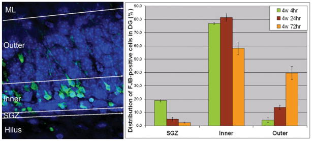
Layer-specific distribution of secondary neuronal death in the hippocampal dentate gyrus following CCI injury. FJB staining to show the degenerating neurons in green in the mouse brain following controlled cortical impact. DAPI was used to stain with DAPI in blue to show the structure of the hilus and the dentate gyrus. Left: Degenerating neurons were observed in the hilus, SGZ, inner granular layer, outer granular layer, and molecular layer (ML). Right: Quantification of degenerating neurons in the hippocampal dentate gyrus. [Color figure can be viewed in the online issue, which is available at www.interscience.wiley.com.]