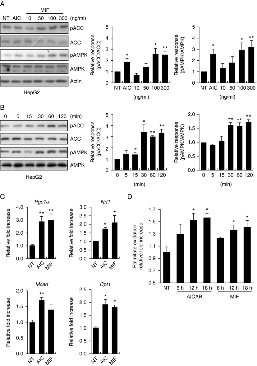
Figure 2.
MIF activates AMPK–ACC, stimulates palmitate oxidation, and increases mitochondria-related gene expression in HepG2 cells. (A) Dose-dependent phosphorylation of AMPK by MIF. HepG2 cells were stimulated with the indicated doses of MIF, AICAR, or vehicle for 1 h. Cell lysates were analyzed by western blotting with anti-phospho-ACC (Ser79) and anti-phospho-AMPK (Thr72) antibodies. Anti-ACC, anti-AMPK, and anti-β-actin antibodies were to check protein loadings. (B) Time-dependent phosphorylation of AMPK by MIF. HepG2 cells were stimulated with MIF (100 ng/μl) for the indicated periods of time. Bar graph depicts the mean (±s.e.m.) ratio of intensity of phospho-ACC-to-total ACC bands and phospho-AMPK-to-total AMPK bands. (C) HepG2 cells were incubated in six-well plates for 24 h with vehicle, MIF (100 ng/μl), or AICAR (100 nM). After 24 h, whole-cell lysates were isolated for analysis of mRNA expression of Pgc1α, Nrf1, Mcad, and Cpt1. (D) HepG2 cells were incubated in 60 mm dishes for 24 h with vehicle, MIF, and AICAR. After washing, cells were assayed for oxidation of [3H]-labeled palmitate, as described in the Materials and methods section. *P<0.05 vs control values (one-way ANOVA). **P<0.01 vs control values. Data are expressed as mean±s.d. of triplicate analyses.

