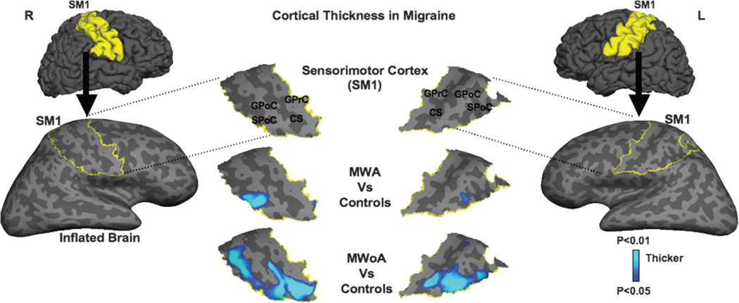
Figure. Mean cortical thickness maps in migraine.
Lateral views of the folded and inflated brain hemispheres exposing sulci (dark gray) and gyri (light gray) with the right and left sensorimotor cortices (SMCs) delineated in yellow (lateral columns). When the migraine subgroups were compared with healthy controls, significant cortical thickening was found in the caudal SMCs of migraineurs, mostly in the somatosensory cortex, where the head is somatotopically represented (center columns). The light–dark blue shading code represents p values for cortical thickness changes. The lighter blue shading indicates thicker cortex. CS = central sulcus; GPoC = gyrus postcentralis; GprC = gyrus precentralis; MWA = migraine with aura; MWoA = migraine without aura; SPoC = sulcus postcentralis.

