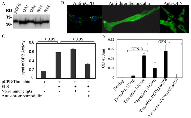
Figure 3. Identification of thrombin-activatable pCPB and its activity in fibroblast-like synoviocytes.
A, Detection of pCPB in OA and RA synovial tissue lysates from two patients each by a monoclonal anti-pCPB antibody in Western blot. B, Immunofluoresence cell labeling of pCPB, thrombomodulin and OPN in FLS. In anti-pCPB panel, green color indicates pCPB and blue color stands for nuclear labeling by DAPI. C, Thrombomodulin expressed on FLS enhanced pCPB activation by thrombin. pCPB activation was blocked in the presence of anti-thrombomodulin antibody. Results represent the mean ± SEM (n = 6). D, Generation of endogenous OPN-R and OPN-L by incubation of thrombin with FLS, with or without exogenous pCPB. Thrombin was used at either 1 U/mL or 10 U/mL; OPN-R or OPN-L in FLS supernatant was measured by direct ELISA as described in Methods. Potato carboxypeptidase inhibitor (CPI) is a specific inhibitor of CPB. Results represent mean ± SEM (n = 6) P < 0.05.

