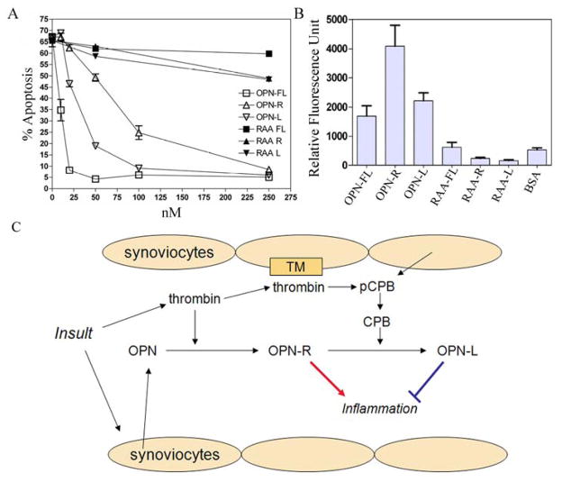
Figure 5. OPN protected neutrophils from apoptosis and fibroblast-like synoviocytes binding to OPN was enhanced in OPN-R but not in OPN-L.
A, Effect of recombinant WT OPN-FL, OPN-R, OPN-L and their RAA substituted counterparts (RAA-FL, RAA-R, and RAA-L) on neutrophil apoptosis. Each data point represents mean ± SEM (n = 3), and the results are representative of one of three separate experiments. B, FLS adhesion to immobilized WT and RGD-substituted OPN-FL, OPN-R, OPN-L. Data are presented as mean ± SEM (n = 8), and results are representative of one of four separate experiments. C, Schematic model of OPN-R and OPN-L in RA: While OPN and pCPB within the inflammatory joint space can be derived from plasma, synoviocytes also produce and release these molecules locally. Following initial insult, thrombin is generated and cleaves OPN to OPN-R, which enhances tissue inflammation. 27 On the other hand, thrombin also binds to thrombomodulin (TM) on the synovial cell surface, which then activates pCPB locally to CPB and converts OPN-R to OPN-L, thereby dampening inflammation.

