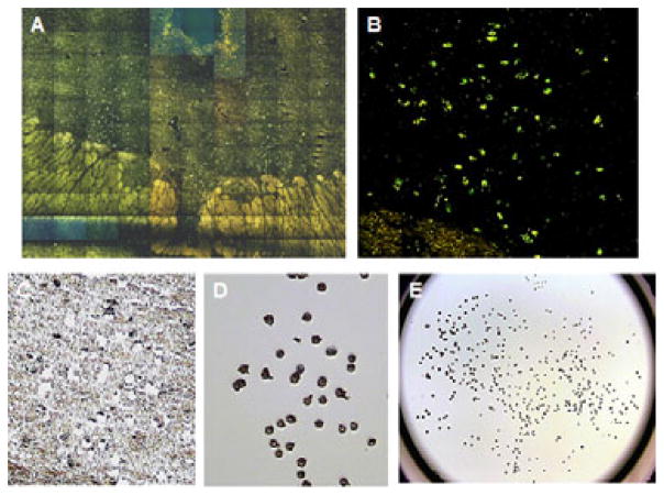
Figure 1.
Representative photomicrographs of dorsal raphe nucleus, immunofluoroscent-stained cells and captured neurons obtained from the laser-capture microdissection instrument. A. High magnification (20x) composite image of entire DR illustrating TPH2-immunofluoroscent stained neurons. B. Photomicrograph of TPH2-immunofluoroscent labeled DR neurons (20x). C. Brightfield photomicrograph of tissue section after the TPH2-stained neurons were captured (20x). D. Photomicrograph of laser-captured serotonin DR neurons adhered to the cap after LCM processing of tissue section in C (20x). E. Photomicrograph illustrating laser-captured serotonin DR neurons adhered to the entire cap after LCM processing of the tissue section.

