
Fig. 2.
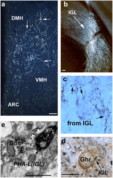
IGL innervation of ghrelin neurons. a–b. The anterograde tracer, PHA-L, was also injected into the IGL (b; bar scale represents 100 μm) of the thalamic LGN. PHA-L-labeled IGL efferents were equally represented in the cell-sparse area of the hypothalamus as were SCN fibers (a; bar scale represents 100 μm). c–d. IGL efferents in this area arborized into putative axon terminals (arrows on c) that were frequently in direct apposition (arrows point to dark boutons on d) to ghrelin-immunoreactive (light brown labeling) cell bodies (bar scale represents 10 μm). e. Electron microscopic analyzes of putative contacts showed symmetrical synaptic contacts between PHA-L-labeled, IGL efferents and immunogold labeled (arrowheads) ghrelin perikarya (bar scale represents 1 μm). oc: optic chiasm; VMH: ventromedial hypothalamic nucleus; DMH: dorsomedial hypothalamic nucleus; ARC: arcuate nucleus.