
Fig. 4.
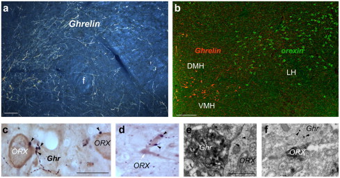
(a) Ghrelin-immunolabeled efferents are abundant in the perifornical region. Bar scale represents 100 μm. (b) Ghrelin-immunoreactive neurons (red fluorescence) are distinct from the lateral hypothalamic orexin/hypocretin neurons (green fluorescence). Bar scale represents 100 μm. (c) In the lateral hypothalamus-perifornical region, ghrelin-immunolabeled axon terminals (arrowheads) are in close proximity to orexin/hypocretin-producing perikarya. Bar scale represents 10 μm. (d) Ghrelin-immunopositive boutons (arrowheads) were also associated with orexin/hypocretin dendrites. (e) Electron micrograph showing direct apposition between a ghrelin-immunoreactive axon terminal and an orexin/hypocretin-labeled (arrowheads point to immunogold) perikaryon. Bar scale represents 1 μm. (f) Electron micrograph showing direct apposition between a ghrelin-immunoreactive axon terminal (arrowheads point to large immunogold particles representing ghrelin immunoreactivity with postembedding labeling) and an orexin/hypocretin-labeled (immunoperoxidase) perikaryon. Bar scale represents 1 μm.