
Fig. 1.
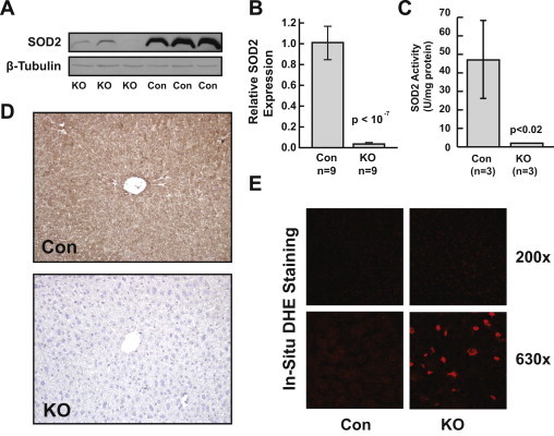
Establishment ofSod2hepatocyte-specific knockout. (A) Western blot for SOD2 demonstrating loss of SOD2 protein in Sod2−/− (KO) livers compared to Sod2L/L (Con) livers. (B and C) Quantification of significant reduction in Sod2 message (B) and SOD2 activity (C) in KO livers compared to control. (D) Immunohistochemical staining for SOD2 demonstrates pan-hepatocellular knockout in Sod2−/− livers compared to control. (E) Representative in situ DHE staining demonstrating enhanced nuclear positivity in Sod2−/− livers compared to control.