
Fig. 4.
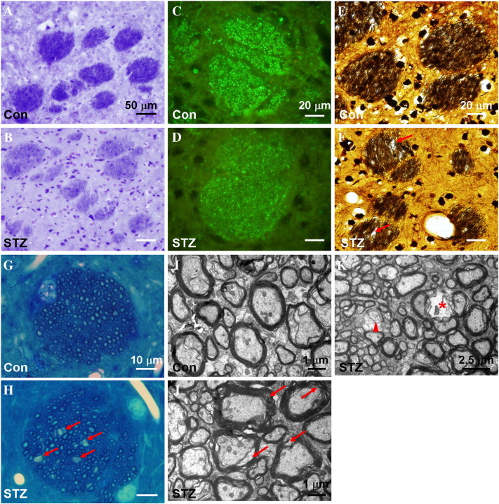
Histopathological staining of the striatal sections from control rats (Con, panels A, C, E, G and I) and STZ-induced rats (STZ, panels B, D, F, H, J and K), including KB staining (A and B), SMI immunohistochemistry (C and D), BS staining (E and F), methylene blue staining (G and H) and uranyl acetate-lead citrate staining (I to K). Compared to that of the control rat, the striatal fiber bundles in the STZ-induced rats are less intensively stained with LFB (B) and SMI-31 (D), and associated with disarranged neurofibrils and vacuole formation (arrows in F and H). Ultrastructural analysis showed that the striatal fiber bundles of the STZ-induced animals are characterized by rarefied myelin sheath (arrows in J), myelin loss (arrow head in K) and signs of axonal degradation (asterisk in K).