Figure 2.

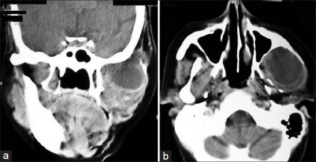
(a and b) Radiological appearance of the lesion in left temporal fossa on computed tomography (Detail description in text)

(a and b) Radiological appearance of the lesion in left temporal fossa on computed tomography (Detail description in text)