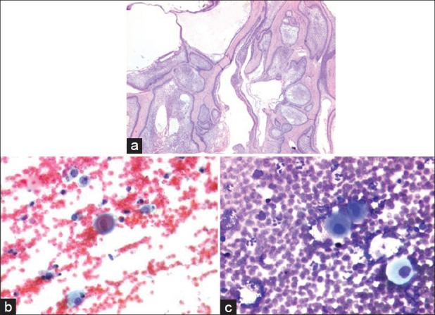
Figure 4.

(a) H and E stained sections showing ameloblastoma with solid and cystic patterns. No squamous differentiation or nuclear atypia was seen (X40). (b and c) Fine needle aspiration cytology of the lesion. Squamous cells showing nuclear atypia admixed with foamy macrophages on a hemorrhagic background. (a) Papanicolaou stain (X400) (b) Giemsa stain (X400)
