Figure 3.

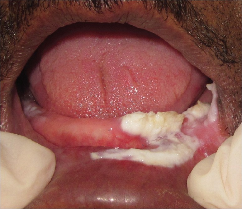
Intra-oral photograph showing lesion in left mandibular alveolus in relation to tooth number 31-35 including vestibule and labial mucosa, crossing the midline

Intra-oral photograph showing lesion in left mandibular alveolus in relation to tooth number 31-35 including vestibule and labial mucosa, crossing the midline