
Figure 5.
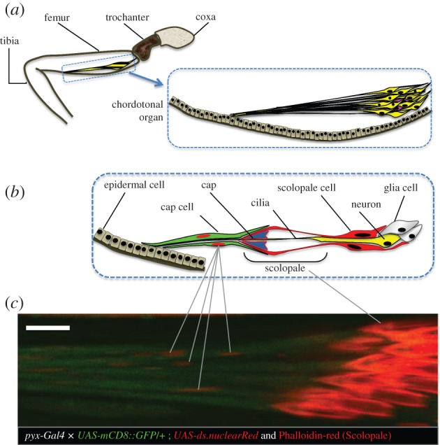
Pyrexia expression in the cho cap cells. (a) Schematic of the general anatomy of the fly's leg with cho neurons indicated in yellow. (b) Detailed representation of the cho showing only one neuron and its associated cells, for simplification purpose. (c) Confocal microscopy of femoral cho of pyx-Gal4 flies expressing mCD8 :: GFP and Nuclear Red. Membrane of Cap cells are shown in green and their nuclei in red. Phalloidin Red was used to stain the scolopales. Scale bar is 10 µm.