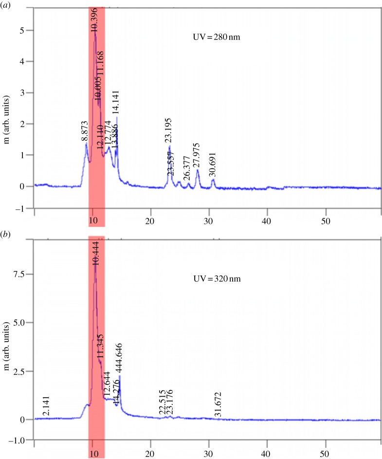
Figure 3.
(a,b) Peaks observed from UV detector of the ivy extract. A prominent peak was observed in both wavelengths (highlighted) during the 10–11 min fraction. This fraction corresponded to the presence of nanoparticles, as indicated by AFM. Peaks with lower intensity were imaged but were found not to contain any nanoparticles. (Online version in colour.)

