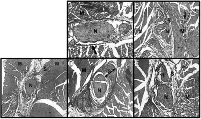
Figure 1.
Photomicrograph of sciatic nerve (N) and surrounding scar (S) and muscles (M) in third week. A: control, B: laceration, C: crush, D: mince and E: burn. In the normal tissue, note the thin darkly stained collagen fibers of epineurium (arrow), which in the scarred tissue becomes a dense band around the nerve (two-head arrow). Masson trichrome staining. (Magnification A: ×100, B-E: ×50

