
Figure 1.
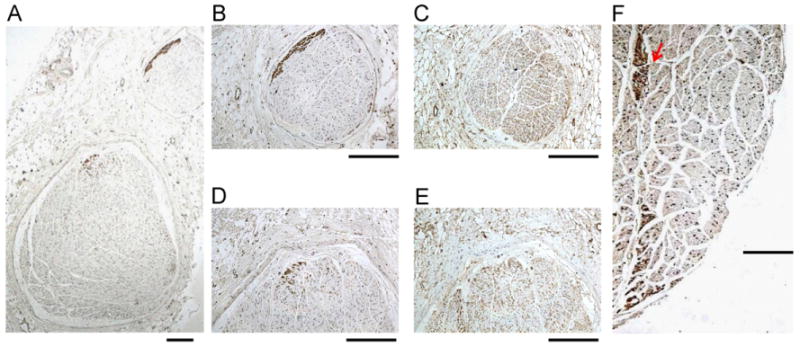
Tyrosine hydroxylase (TH) and cholineacetyltransferase (ChAT) staining of the cervical vagus nerves (CVNs). A: A low power view of the right CVN stained with TH. There are 2 distinct nerve bundles in this nerve. B, D: The TH stain of the smaller (B) and the larger (D) bundles. The brown color identifies the positively stained nerves. Note that TH-positive nerves are located in the periphery of the nerve bundle. C, E: ChAT staining of the same structures as in panels B and D, respectively. Note that ChAT-positive components are widely distributed in the CVN. F: The TH-positive nerve structure (red arrow) in the middle of the CVN. The objective lens used in panel A was 4×, with a calibration bar of 0.2 mm in length. The objective lens used in panels B–F was 20×, with a calibration bar of 0.2 mm in length.