
Figure 4.
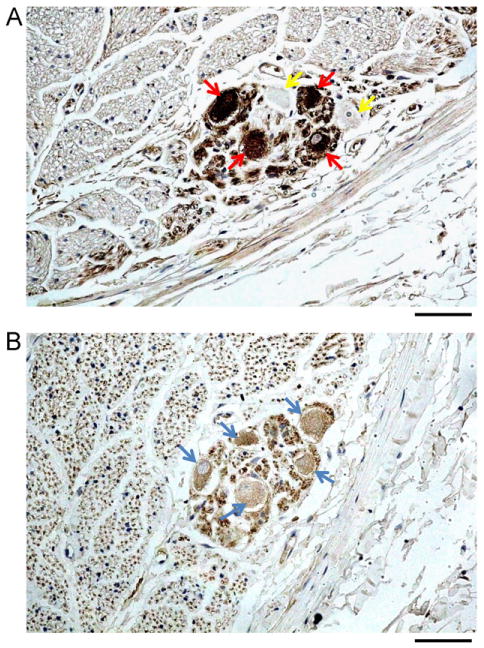
Tyrosine hydroxylase (TH) and cholineacetyltransferase (ChAT) staining of an autonomic ganglion in the left cervical vagus nerve (CVN). A: Four ganglion cells stained positive for TH (red arrows) and 2 stained negative (yellow arrows). B: The same nerve with 5 ganglia stained ChAT positive (blue arrows). The objective lens in panels A and B was 40×, with a calibration bar of 0.05 mm in length.