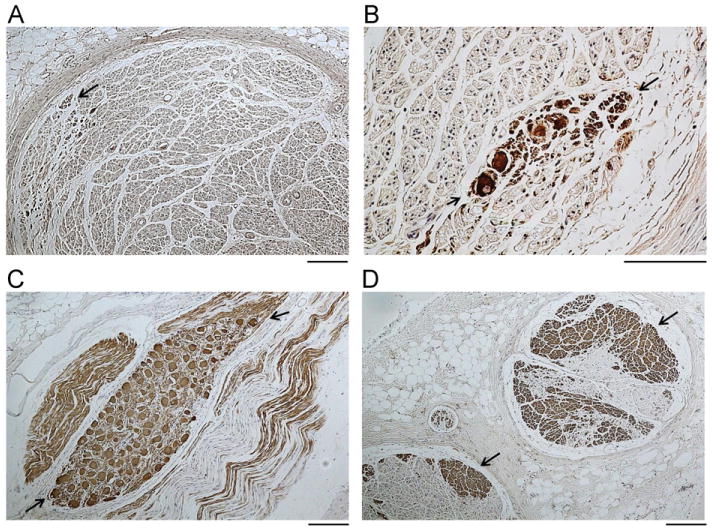
Figure 6.

Tyrosine hydroxylase (TH) staining of the cervical (A and B) and thoracic (C and D) vagal nerves from the same dog as in Figure 5. Positive stains are brown (arrows). The TH-positive nerves in cervical vagal nerves are located in the periphery (A), while abundant TH-positive nerves are present in the thoracic vagal nerve both in the periphery and in the center (D). Note that TH-positive ganglion cells (between arrows) are present both in the cervical vagal nerve (B) and in the thoracic vagal nerve (C). The objective lens is 10× in panels A, C, and D and 40× in panel B. The calibration bars in panels A, C, and D are of 0.1 mm in length. The one in panel B is of 0.05 mm in length.
