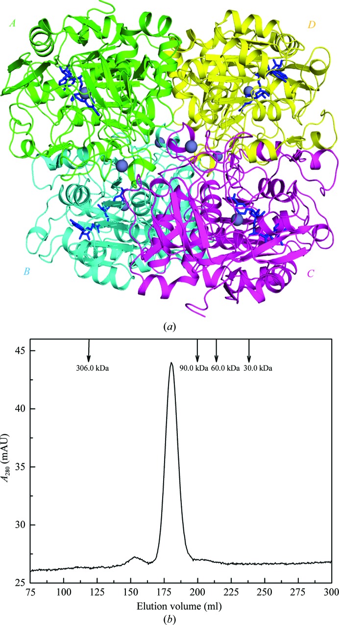
Figure 1.
Tetrameric NAD+-bound formaldehyde dehydrogenase (FDH) from P. aeruginosa. (a) The overall structure of the FDH tetramer. Four subunits (A–D) are shown in ribbon mode in different colours. The zinc ions are depicted as grey spheres and the bound NAD+ ions are shown as blue sticks for each of the four subunits. (b) SEC analysis of the FDH at a concentration of 2 mg ml−1. The protein sample was loaded onto a HiLoad 26/60 Superdex 200 column and was eluted at a flow rate of 3 ml min−1 with detection of the absorbance at 280 nm. The elution profiles of four molecular-mass protein standards are also shown as labelled arrows.

