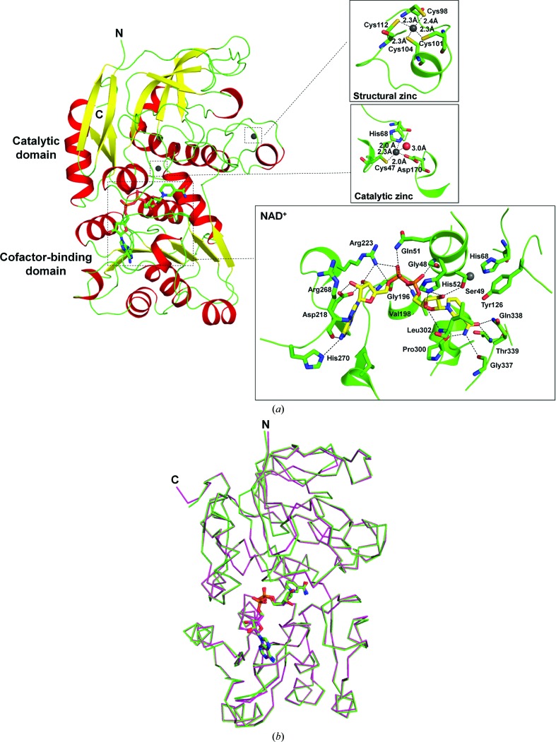
Figure 2.
The structure of the P. aeruginosa FDH subunit. (a) Ribbon drawing of FDH subunit A. The N- and C-termini are labelled by the letters N and C, respectively. α-Helices are coloured red, β-strands yellow and loops green. The NAD+ and zinc ions are highlighted as sticks and spheres, respectively. The insets show the binding modes of two zinc ions (grey spheres) and the cofactor NAD+ (sticks) to the FDH subunit. Hydrogen bonds are indicated by dotted lines. The residues of FDH involved in binding are shown as sticks and the residue numbers are indicated. (b) Superimposition of FDH subunit A (green) with a subunit of P. putida FDH (magenta). Proteins are indicated by Cα traces with N- and C-termini marked and the bound NAD+ ions are shown in stick representation.

