
Figure 1.
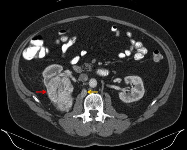
Diagnostic contrast-enhanced computed tomography (CT) of the abdomen (axial view) showing a solid mass in the superior pole of the right kidney (red arrow). This lesion demonstrated contrast enhancement with delayed washout having a density prior to intravenous contrast of 30 Hounsfield units (HU), 150 HU after contrast administration, and 58 HU in the excretion phase, suspicious for clear cell renal cell carcinoma. In retrospect, the small retroperitoneal lymph node (yellow arrow), which was not originally identified in the initial interpretation of the prior contrast-enhanced CT of the abdomen or the 18F-fluorodeoxyglucose positron emission tomography (PET)/CT, was later first identified on 124I-labeled chimeric monoclonal antibody G250 PET/CT and proven to represent a retroperitoneal lymph node metastasis by light microscopy.