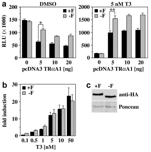
Fig. 3. The F-domain does not regulate the hormone responsiveness of TRαA in mammalian cells.

A, Luciferase activity (RLU) of CV1 cells transiently transfected with DR4 luciferase and CMV β-galactosidase reporters and various amounts of either pcDNA3 TRαA1 (+F) or pcDNA3 TRαA1-F (−F). Cells were treated with either DMSO (vehicle) or 5 nM T3. Shown are the averages and standard deviation of 3 independent experiments performed in triplicate. * A student’s t test shows that the difference in the activities of TRαA1 and TRαA1-F is statistically significant (p<0.001). B, Dose response analysis of the activity of TRαA1 and TRαA1-F (5 ng each) in the presence of various T3 concentrations (0.1 - 50 nM). Fold induction is defined as RLU(+H)/RLU(−H). Shown are the averages and standard deviations of three independent experiments performed in triplicate. C, Expression of TRαA1 and TRαA1-F in transiently transfected CV1 cells (30 μg total protein) monitored with a HA-tag specific antibody. Equal loading and transfer of CV1 expressed TRs were controlled by Panceau Red-staining of the blot. A selected Panceau Red-stained protein band is shown as loading control.
