
Fig. 4.
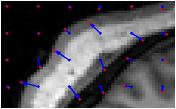
Registration of brain MRI slices: close-up on the left posterior area of the source image. Initial control points in blue moved to the positions shown in red. They moved toward the contours of the image. They do not move in absence of image force in homogeneous areas. σ = 5 voxels and γ = 10−3.