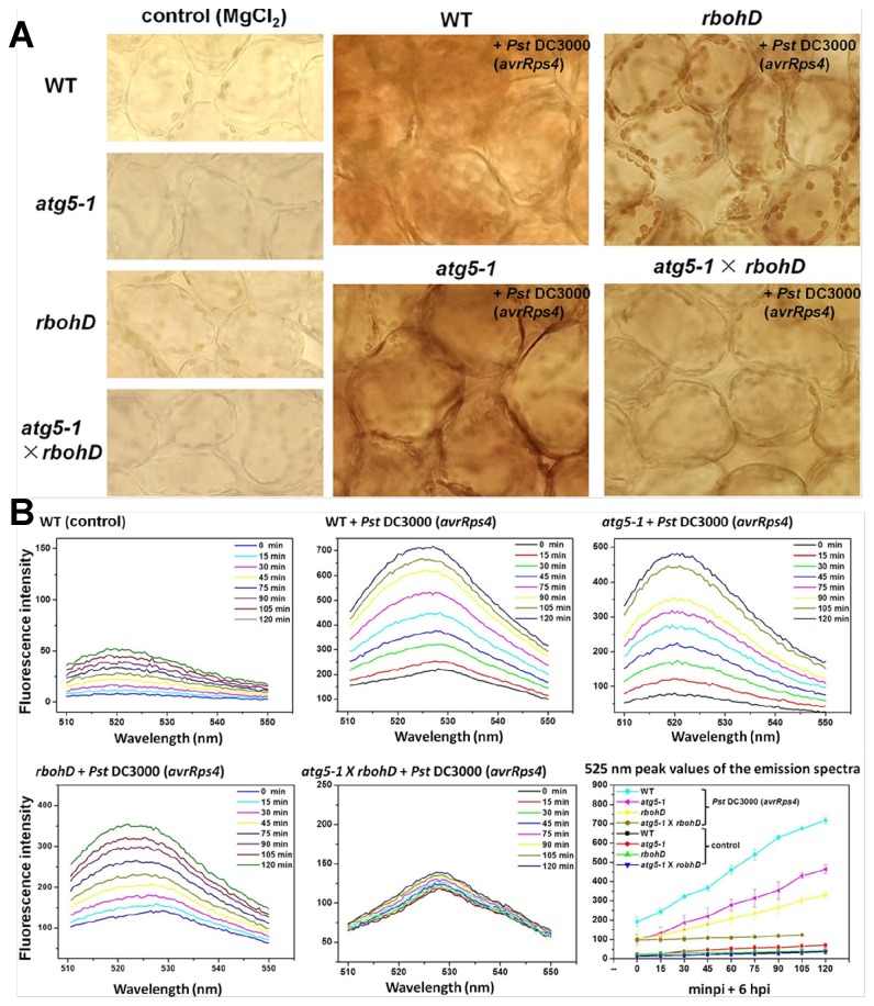
Figure 8. The accumulations of H2O2 induced by pathogens in WT, atg5-1, rbohD and atg5-1 × rbohD.
A. The plants (4 weeks old) were infiltrated with Pst DC3000 (AvrRps4) (OD600 = 0.2) or MgCl2 (control). DAB staining of leaves from WT, atg5-1, rbohD and atg5-1 × rbohD were taken after 24 hpi, respectively. Experiments were performed three times with similar results. B. Show are Arabidopsis leaves after infiltrating Pst DC3000 (AvrRps4) (OD600 = 0.2) or 10 mM MgCl2 (control) for 6 hpi. Then the changes of 525 nm peak values in fluorescence emission spectra were scanned for 120 min. Excitation wavelength: 488 nm; Excitation slit width: 10 nm; Emission slit width: 8.5 nm; Scanning speed: 200 nm/min; Scanning wavelength range: 510-550 nm.

