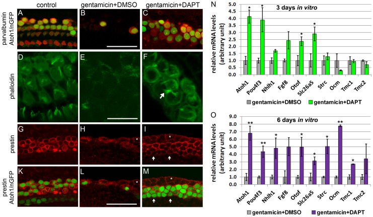
Figure 4. Newly generated hair cell-like cells mature in vitro.
Hair cell phenotype of untreated (control A, D, G, K) gentamicin +DMSO (B, E, H, L) and gentamicin +DAPT (C, F, I, M) treated cochlear explants. A–C: Parvalbumin immuno-staining (red) and native Atoh1/nGFP (green) expression mark hair cells. After 3 DIV, Atoh1/nGFP positive cells (green nuclei) in gentamicin + DAPT treated (C) cultures express hair cell marker parvalbumin (red). Scale bar 50 μm. D–F: Fluorescently labeled phalloidin (green) visualizes actin. After a total of 3 DIV, gentamicin + DAPT (F) treated cultures have actin-rich (phalloidin, green) membrane protrusions (white arrows) reminiscent of hair cell stereocilia. Scale bar 25 μm. G–M: Prestin immuno-staining (red) selectively marks outer hair cells. After 6 DIV, the majority of Atoh1/nGFP positive cells co-express prestin in gentamicin + DAPT treated cultures (I, M). Note white asterisk labels Atoh1/nGFP negative epithelial cells at the lateral edge of cochlear epithelium, which express prestin at low levels (H, I, L, M). Scale bar 50 μm. N–O: Q-PCR based quantification of mRNA expression levels of hair cell specific genes in hair cell damaged (gentamicin +DMSO) and hair cell damaged and Notch inhibited (gentamicin +DAPT) cochlear epithelia after 3 DIV (N) and 6 DIV (O). Data expressed as mean ± SEM, (n = 3 independent experiments, *p≤0.05, Student's t-test).

