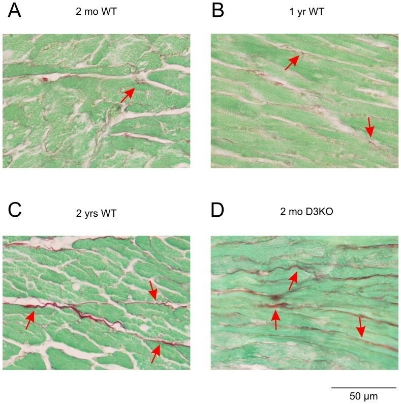
Figure 4. Myocyte interstitial fibrosis with age.
Representative images of the left ventricle myocardium of 2 mo old WT (A), 1 yr old WT (B), 2 yr old WT (C), and 2 mo old D3KO mice (D). Images show increased content and thickness of fibrillar collagen with age (arrows) in WT hearts and in young D3KO hearts.

