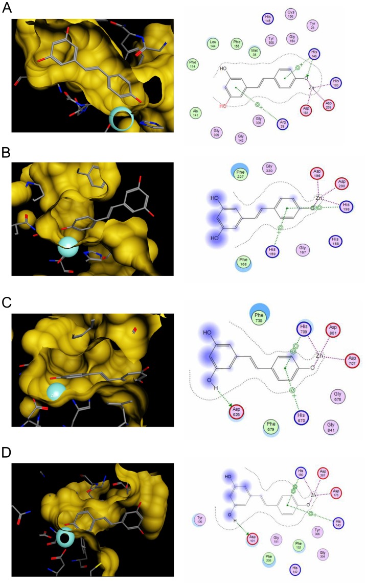
Figure 1. In silico docking analysis with resveratrol illustrates an inhibitory potential for human HDAC enzymes of classes I and II.
(A–D) Results of the in silico docking analysis of resveratrol with crystal structures of HDAC2 (A), HDAC4 (B), HDAC7 (C) and HDAC8 (D). The analysis demonstrates the predicted binding mode of resveratrol in the different HDAC binding pockets. The predicted interactions of resveratrol with the zinc ion (turquoise sphere) and other residues of the catalytic center are highlighted in the left row. 2D depiction of resveratrol along with interacting amino acids is shown in the right row. Green circles represent hydrophobic, purple circles polar, red circles acidic and blue circles basic residues. Blue halolike discs around amino acids are calculated based on the reduction of solvent exposure by the ligand. Blue arrows represent backbone H-bond interactions, green ones depict sidechain H-bond interactions. Green benzol rings with a “+” describe arene-cation interaction, 2 benzol rings an arene-arene interaction. Areas with a blue background are solvent exposed parts of the ligand. The purple dotted lines represent metal contact.

