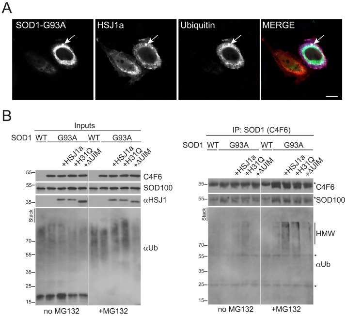
Figure 9. hHSJ1a enhances SOD1G93A ubiquitylation.
(A) Representative immunofluorescence image showing co-localization of mutant GFP-SOD1 (SOD1-G93A, green), myc-HSJ1a (S653, red) and myc-ubiquitin (pan Ub, blue) within an intracellular inclusion (arrowed) in SK-N-SH cells, following co-transfection and proteasome inhibition with MG132. Scale bar: 10 µm. (B) Western blots with anti-SOD1 (C4F6 or SOD100), anti-HSJ1 (S653) and pan-ubiquitin (Ub) antibody as indicated, showing input and C4F6-immunoprecipitated material from SK-N-SH cells transfected and treated with MG132 as indicated. An increase of ubiquitin reactivity of in the presence of WT HSJ1a was observed upon proteasome inhibition. The HSJ1a H31Q mutant did not stimulate proteasomal degradation of ubiquitylated SOD1, whereas the HSJ1a ΔUIM mutant did not stimulate SOD1-G93A ubiquitylation. Asterisks indicate IgG bands. The position of molecular weight markers is indicated on the left.

