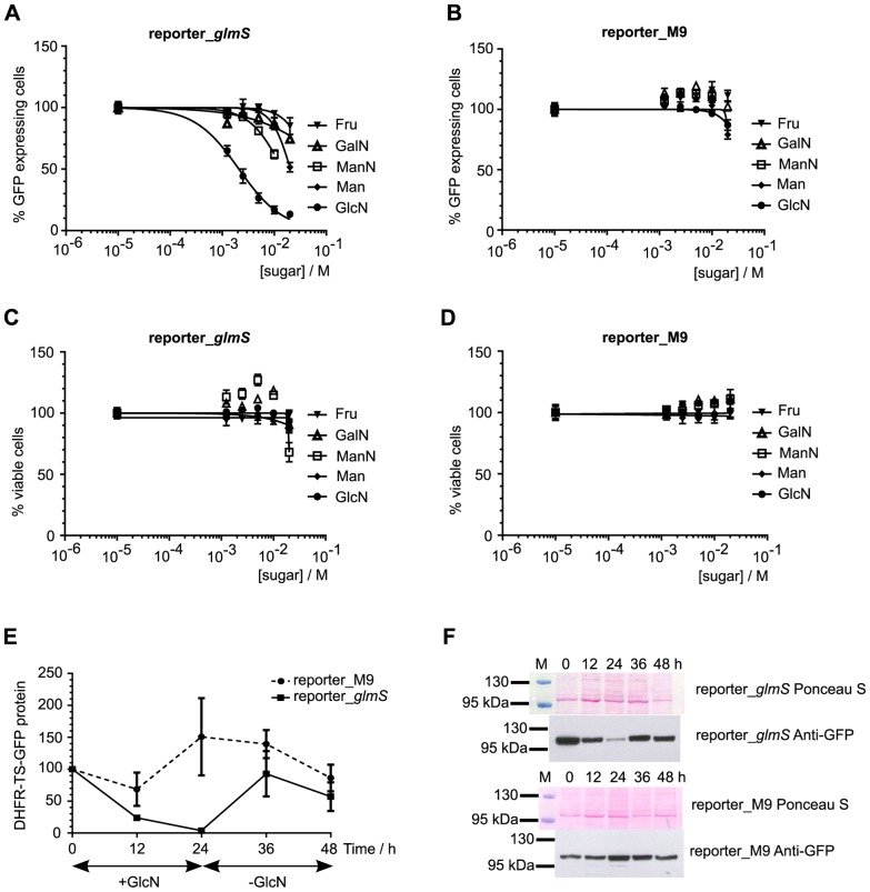
Figure 3. Ribozyme-mediated knockdown of reporter protein expression in response to sugar treatment.
Ring-stage synchronized parasites from reporter_glmS (parts A, C) and reporter_M9 (parts B, D) transfected lines were treated for 24 h with varying concentrations of different sugars (GlcN, ManN, GalN, Fru and Man) prior to flow cytometric analysis. The numbers of GFP positive and viable cells were normalized to untreated controls, which were set as 100%. Data are the mean from triplicate experiments and error bars represent S.E.M. Part E shows that ribozyme-mediated knockdown of reporter protein is reversible. 10% ring-stage parasites were treated with 10 mM GlcN for 24 h and samples taken after 12 and 24 h of treatment. The parasite culture medium was changed and the culture was continued, with samples taken after 12 and 24 h in the new GlcN-free medium. The experiment was performed in triplicate with independent cultures for each replicate (5 sampled time points from each culture). Western immunoblot analysis of DHFR-TS-GFP reporter protein expression was performed using an anti-GFP polyclonal antibody. The normalized intensity of the ∼100 kDa band corresponding to DHFR-TS-GFP protein was measured relative to the normalized intensity at 0 h, which was taken as 100%. Data are the mean and error bars represent S.E.M. Representative images from Ponceau S staining of parasite lysates following electrophoresis and transfer to membrane, and the corresponding chemiluminescent detection of GFP with specific antibodies are shown in part F. The pre-stained marker lane is marked M above the Ponceau S panel, and the sizes of two marker proteins are indicated.

