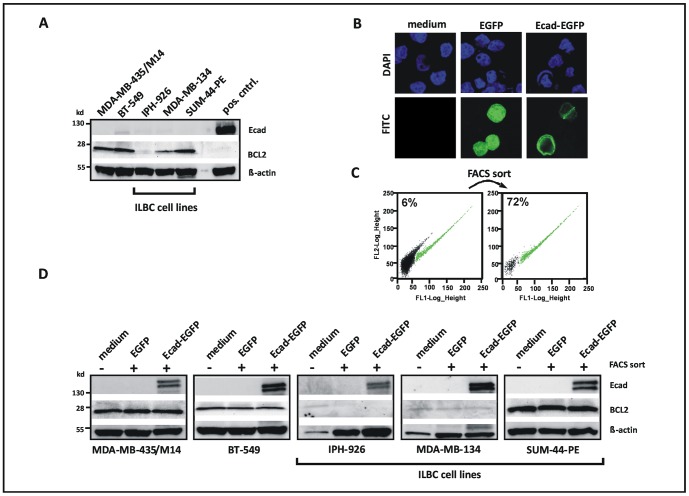
Figure 1. Reconstitution of E-cadherin fails to impact on BCL2 expression in human ILBC cells.
(A) Analysis of E-cadherin (Ecad) and BCL2 protein expression by Western Blot. Pos cntrl; positive control for E-cadherin. (B) Fluorescent imaging of cells subjected to ectopic expression of EGFP or EGFP-tagged E-cadherin (Ecad-EGFP) by transient transfection. Representative photomicrographs show the IPH-926 ILBC cell line. Note the membranous localization of Ecad-EGFP. (C) Pre-analytical enrichment of EGFP- or Ecad-EGFP-positive cells by FACS sort. Cells within the EGFP-positive gate are colored in green. Representative dot blots show IPH-926 ILBC cells transfected with the Ecad-EGFP expression construct. (D) Analysis of E-cadherin and BCL2 protein expression by Western blot. Cells were subjected to ectopic expression of EGFP or Ecad-EGFP for 24 h and subsequent pre-analytical enrichment of EGFP-positive cells by FACS sort (minimum 70% purity). Untreated cells were included as controls. Ecad-EGFP presented as a double band of approximately 150 kd, as reported previously [27]. Similar results were obtained 48 h after transfection (not shown).

