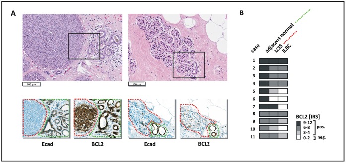
Figure 4. Similar or reduced BCL2 immunoreactivity in LCIS compared with the adjacent mammary epithelium.
(A) Representative photomicrographs showing two LCIS lesions characterized by either similar (left, case #1) or reduced (right, case #10) BCL2 immunoreactivity compared with the adjacent E-cadherin-positive epithelium. HE stained sections are shown on top. Serial sections subjected to immunohistochemical staining of E-cadherin (Ecad) and BCL2 are shown below. (B) Overview on BCL2 immunoreactivity in LCIS lesions of n = 11 patients. At least three TDLUs affected by LCIS were considered per case, but showed essentially the same staining characteristics (not shown). A color scale indicative of the IRS is included on the right side.

