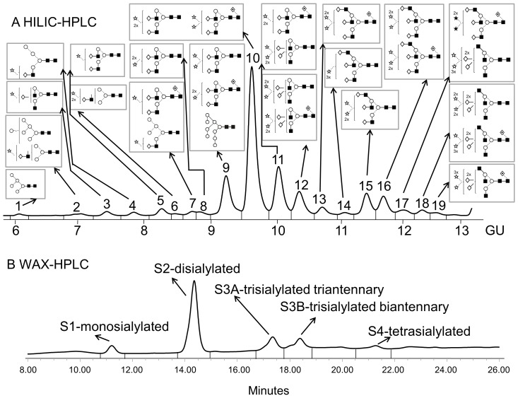
Figure 2. Typical HILIC- (A) and WAX-HPLC (B) chromatograms of mouse serum N-glycans.
Structural assignments are in Table 1 and Figure S1. The HILIC-chromatogram was separated into 19 peaks and the WAX-chromatogram was separated into 5 peaks: S1, S2, S3A, S3B and S4. Symbols encode the following monosaccharide structures: GlcNAc, filled square; mannose, open circle; galactose, open diamond; fucose, diamond with a dot inside; Neu5Gc sialic acid, star with dot inside; beta linkage, solid line; alpha linkage, dotted line (Harvey et al [36]).

