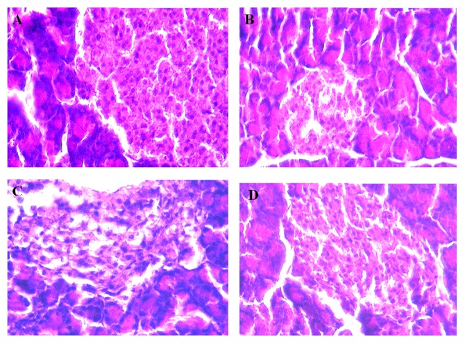
Figure 6. Photomicrographs of histological changes of rat pancreata (Gomori stain).

A) Non-diabetic: Normal histological structure of rat pancreata showing average sized islets (red arrow) and normal sized β cells. B) Diabetic control rat pancreata showing β cells slightly elongated with more destruction (++++). C) Concomitant administration of sitagliptin (5 mg/kg, p.o.) with cycloart-23-ene-3β, 25-diol (1 mg/kg, p.o.) treated rat pancreata showing slightly elongated of the β cells with less destruction (+). D) Concomitant administration of sitagliptin (5 mg/kg, p.o.) with L-glutamine (1000 mg/kg, p.o.) treated rat pancreata showing slightly elongated of the β cells with less destruction (+). Grade: – No injury; Grade: ++++ severe injury; Grade: ++ mild injury; Grade: + Very mild injury.
