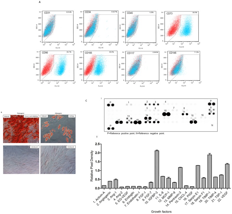
Figure 1. Characterization of rat adipose-derived stem cells (rADSCs).
(A) FACS analysis of cultured rADSCs (p3) showed that surface antigens expression of rADSCs consistent with mesenchymal stem cell markers. Plots show the isotype control (red) versus specific antibody staining (blue). (B) Photomicrograph of osteogenic induced rADSCs (left; Alizarin red S staining) and adipogenic induced rADSCs (right; Oil Red O staining). Scale bar = 50 µm. (C) Proteomic profile angiogenesis array analysis of human ADSCs showed that 22 angiogenesis-related proteins were detected in the supernatant of human ADSCs.

