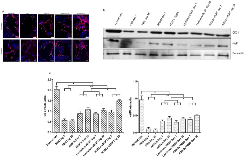
Figure 6. ADSCs-VEGF restored cavernous endothelial content in STZ-induced diabetic rats.
A: Immunofluorescent staining of cavernous tissue using CD31 and vWF antibodies (showed by white arrow) in PBS-treated STZ-induced diabetic rats or ADSCs or lentivirus-VEGF or ADSCs-VEGF treated DED rats and normal rats. Scale bar = 50 µm. B: Immunoblotting analysis of the CD31 and vWF in the normal rats and STZ-induced diabetic rats after 1 weeks or 4 weeks treatment of PBS or ADSCs or lentivirus-VEGF or ADSCs-VEGF. C: Quantitative analysis of the immunoblotting by using Quantity One® software. *p<0.05, **p<0.01.

