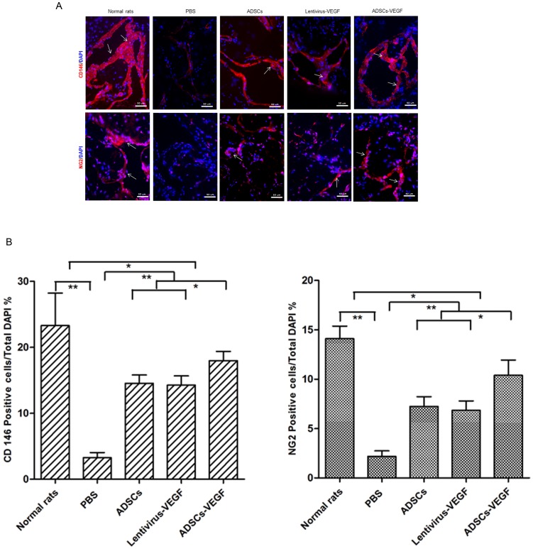
Figure 8. ADSCs-VEGF ameliorated pericytes markers in the cavernous tissue of DED rats.
Immunofluorescent staining (A) and hemi-quantitative analysis (B) showed decreased pericytes markers (CD146, NG2) in the penis of STZ-induced DED rats treated by PBS compared with age-matched normal rats. And ADSCs or lentivirus-VEGF treatment partly but ADSCs-VEGF showed more effectively recovery of these markers. Scale bar = 50 um; *p<0.05, **p<0.01.

