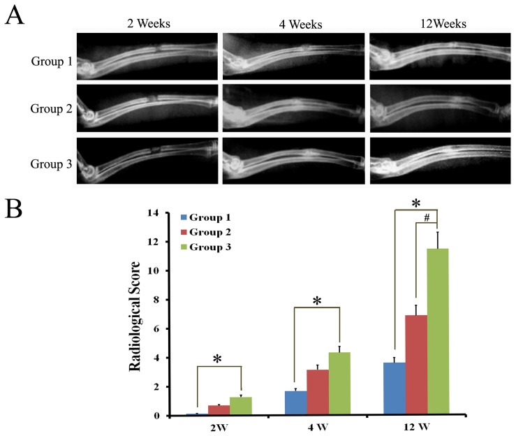
Figure 7. Radiography observation in bone defect model.
A) X-rays photograph of rat radical defects in different groups at 2 weeks, 4 weeks, and 12 weeks showing complete bridging of the defect in group 3, however, a radiolucent line was still present at the defect site in group 1 at 12 weeks. B) Radiographical scoring also showed that new bone formation in group 3 was more significant difference than the other groups at 12 weeks (p<0.05). (Group 1 referred to β-TCP group; Group 2 referred to ADSCs/β-TC group; Group 3 referred to Ad-CGRP-ADSCs/β-TCP group).

