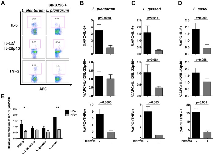
Figure 5. Enhanced APC inflammatory response to commensal lactobacilli signals predominantly through p38-MAPK.
(A) Representative flow cytometry plot and frequencies of APCs from HIV-infected patients producing proinflammatory cytokines IL-6, IL-12/IL-23p40, and TNFα in response to (B) L. plantarum WCFS1 (n = 16), (C) L. gasseri 1SL4 (n = 12), and (D) L. casei BL23 (n = 11) with or without BIRB796 pretreatment. (E) Relative expression of MKP-1 in unstimulated and bacterial stimulated APCs as determined by real-time PCR (HIV− n = 3–9, HIV+ n = 5–16). Bar graphs represent mean +/− SEM. P values determined using paired t or Mann Whitney U test (*P<0.05, **P<0.01, ***P<0.001).

