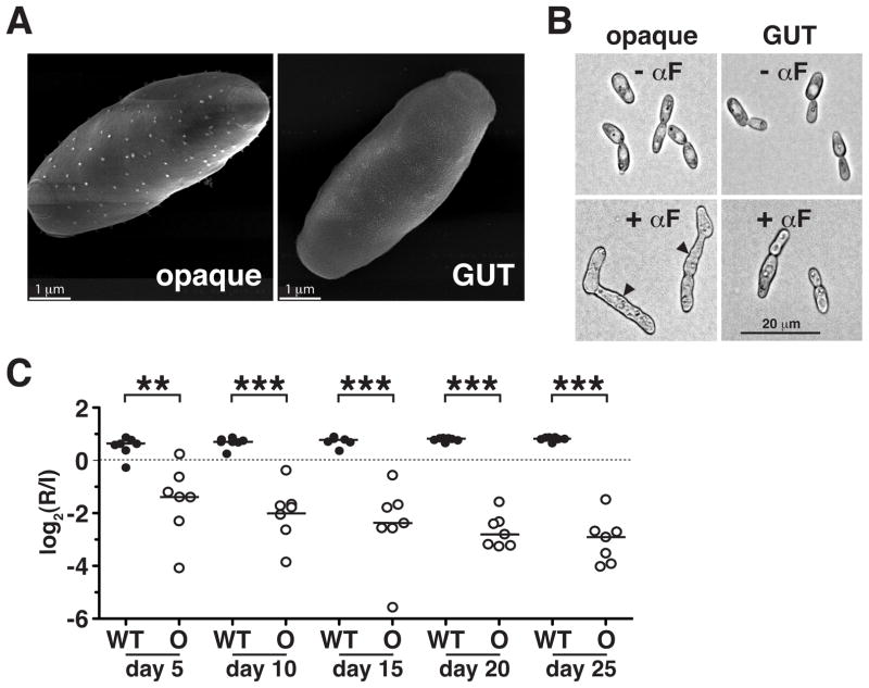
Figure 3. GUT cells are distinct from previously identified opaque cells.
A) Scanning electron micrographs showing “pimples” on the surface of opaque (SN967) but not GUT (SN1045) cells. B) Opaque but not GUT cells form mating filaments in response to mating pheromone (αF). Arrowheads indicate mating projections. All images were obtained at the same magnification. C) Opaque cells (SN967) are significantly outcompeted by wild type (SN425) in the murine commensal model (n = 7 animals). ** p<0.005, *** p<0.001 by the t-test. A replicate of this experiment yielded similar results (Pande and Noble, unpublished).

