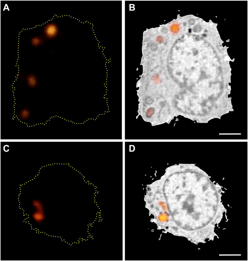
Figure 4.
Correlated lymphocyte Fluorescently Stained with LysoTracker. (A and B) Lymphocytes were fluorescently labeled with LysoTracker vital dye. A through-focus fluorescence dataset was collected using a cryo confocal light microscope. Weak non-specific Lysotracker staining was present throughout the cell, which was used to align the fluorescence and SXT reconstructions. Several round organelles were observed in the SXT reconstruction that correlated with the punctate Lysotracker fluorescence. Scale bar = 2 μm.

