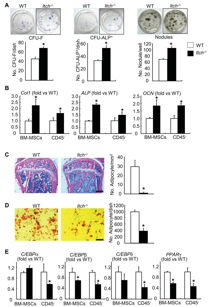
Figure 2. Itch−/− bone marrow mesenchymal progenitors have increased osteoblast differentiation and decreased adipocyte differentiation.
(A) Bone marrow mesenchymal progenitor cells (BM-MPCs) from Itch−/− mice and WT littermates were cultured in the osteoblast-inducing medium for 7-21 days. (A) Representative image of CFU-F, CFU-ALP+ colonies or mineralized nodules (upper panels) and the number of CFU-F and CFU-ALP+ colonies or mineralized nodules (lower panels). Values are mean ± of 3 dishes. (B) The expression levels of osteoblast marker genes were examined by qPCR in B-MPCs and CD45− MPC-enriched cells. (C) H&E-stained sections from tibial bone of 3-month-old Itch−/− mice and WT littermates were analyzed. The number of adipocytes/mm bone surface was counted. Bar = 250μm. Values are mean ± SD of 5 mice/group. (D) BM-MPCs were cultured in the adipocyte inducing medium for 21 days and the number of adipocytes was counted. Bar = 100μm. Values are mean ± SD of 3 dishes. (E) The expression levels of adipocyte master genes were examined by qPCR in BM-MPCs and CD45− MPC-enriched cells. Values are mean ± SD of 5 mice. *p<0.05 vs WT cells.

