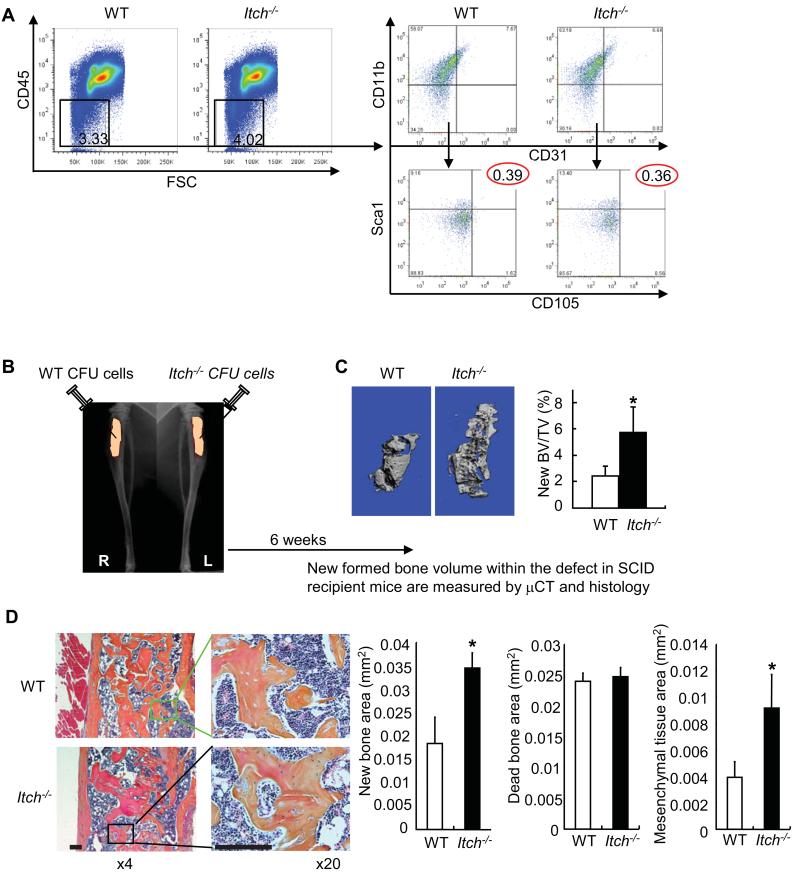
Figure 3. Mesenchymal colony cells from Itch−/− mice form increased new bones in recipient mice.
(A) The % of non-hematopoietic lineage cells (CD45−) and the % cells that express MPC surface markers (CD11b−/CD31−/Sca1+/CD105+) in primary bone marrow cells by FACS analysis. (B) CFU cells from Itch−/− and WT littermates were implanted into tibial defects of SCID mice with decalcified bone matrix as scaffold. Mice were sacrificed 6 weeks post-implantation. (C) μCT images and μCT data of the % of new formed bone volume vs total tissue volume. (D) Histology and histomorphometric data of the area of new bone, dead bone and mesenchymal tissue containing spindle-shaped fibroblasts observed in decalcified H&E-stained sections of the bones Bar = 125μm. Values are mean ± SD of 5 mice. *p<0.05 vs WT cells.

