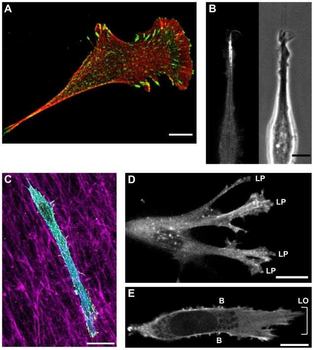
Figure 2. Mode of cellular protrusion as determined by ECM dimensionality.
A) NIH/3T3 fibroblast demonstrating a classic hand-mirror morphology on a 2D substrate; red is phalloidin staining and green shows activated β1 integrin adhesions. B) eGFP-VASP (left) and phase contrast (right) image of a NIH/3T3 fibroblast migrating along a 1D micropatterned line. C) NIH/3T3 fibroblast within a 3D-CDM showing staining for F-actin (phalloidin, cyan), paxillin (yellow), and fibronectin (magenta). D). eGFP-actin expressed in a human foreskin fibroblast illustrating lamellipodia in non-linear 3D collagen. E) Lobopodia (LO) and lateral blebs (B) shown by eGFP-actin as a human foreskin fibroblast migrates through a linear-elastic 3D-CDM. Scale bars: 10 μm.

