
Figure 1.
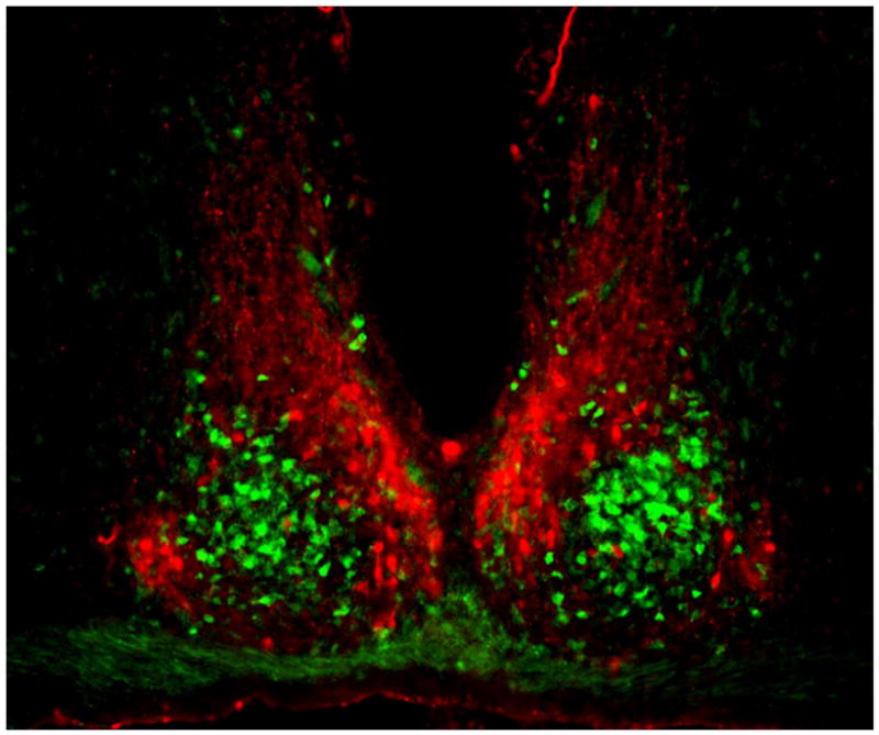
Coronal section of mouse SCN, showing the ventral core region delineated by green fluorescent protein (GFP) expressed in GRP neurons (green) and the dorsal shell region delineated by immunofluorescent labeling for AVP (red). Between left and right SCN is the third ventricle, and below is the optic chiasm. Reprinted from Karatsoreos et al. (185), with permission pending from the Society for Neuroscience.