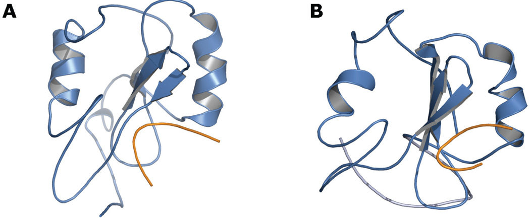
FIGURE 2.
Molecular dynamics snapshots of the Grb7-SH2 domain (blue) in complex with the erbB2 receptor peptide pY1139 (orange). A, Integrity of the canonical SH2 domain fold following 21 nanoseconds of unrestrained explicit solvent molecular dynamics with a 1-femtosecond time step. B, Destructuring of the domain’s C-terminal region during explicit solvent simulations with a 2-femtosecond time step and with higher average total and potential energy than the simulation represented in panel A. The image shown is a snapshot taken after 11 nanoseconds of unrestrained dynamics at 300 K.

