Figure 6.
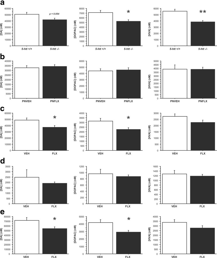
5-HTT blockade reduces DA and DA metabolite levels in frontostriatal regions. a–e, DA, DOPAC, and HVA levels were measured by high-performance liquid chromatography in 5-htt−/− mice (a), after transient developmental 5-HTT blockade (FLX; 10 mg/kg/d, i.p.; P4–P21; b), and after chronic fluoxetine treatment (FLX; 10 mg/kg/d; drinking water; c–e). Total frontostriatal (a–c), frontal cortex (d), and dorsal striatal (e) levels were assessed. DOPAC and HVA levels, normalized by tissue weight, decrease after constitutive genetic 5-htt ablation (a). No changes of DA, DOPAC, or HVA levels, normalized by tissue weight, were detected after transient developmental 5-HTT blockade (b). DA and DOPAC levels, normalized by tissue weight, decrease after chronic FLX treatment in frontal–striatal (c) and dorsal–striatal (e) tissue samples. n = 6–13 mice per group. *p < 0.05; **p < 0.01.

