
Figure 2.
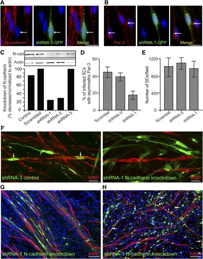
Knockdown of SC N-cadherin disrupts the asymmetric localization of Par-3 and delays the initiation of myelination. A, SC–DRG cocultures were infected with retroviral vectors coexpressing GFP and the shRNA for N-cadherin. N-cadherin is detected by immunostaining (red), and SCs infected with shRNA-1 are identified by the expression of GFP (green). The arrow indicates an infected SC and the corresponding knockdown of N-cadherin. B, SC–DRG cocultures were infected with shRNA-1 against N-cadherin and stained for Par-3 (red). The arrows indicate uninfected SCs displaying asymmetric localization of Par-3, whereas Par-3 localization is disrupted in the SC expressing GFP. Scale bars, 10 μm. C, To determine the efficiency of knockdown with shRNA, purified SCs were infected with a control (GFP only), a scrambled shRNA, shRNA-1, -2, and -3. Cells were extracted and analyzed by Western blot analysis. Quantification of the Western blot is displayed as a percentage decrease normalized to β-actin. D, SCs infected with scrambled shRNA, shRNA-3 (control), and shRNA-1 against N-cadherin were quantified as a percentage of infected SCs that display asymmetric Par-3. Knockdown of N-cadherin using shRNA-1 results in a twofold decrease in the number of infected SCs displaying asymmetric Par-3. E, Manipulation of N-cadherin expression does not have an appreciable effect on SC proliferation, as determined by the number of SCs per image field. Error bars indicate SD. F, G, Using SC–DRG cocultures, SCs were infected with either shRNA-3 (control) or shRNA-1 against N-cadherin and immunostained for MBP (red) 10 d after induction of myelination. Infected cells were indicated by the concomitant expression of GFP (green). The arrows indicate myelinating SCs expressing the control shRNA. SCs infected with shRNA-1 against N-cadherin did not form myelin as detected by MBP expression (red). Scale bars: F, 10 μm; G, H, 50 μm. H, After 15 d of induction of myelination, SCs infected with shRNA-1 were indistinguishable from the control cultures and were myelinating axons in similar proportions (red). The arrows indicate cells expressing GFP (green) and MBP (red), indicating that the knockdown of N-cadherin in SCs delays but does not inhibit the initiation of myelination.